The various control functions of the brain are often implemented in networks of cortical and subcortical areas distributed across the brain. Therefore, to understand the neural underpinnings of these control functions, we must do more than understand how the neurons in one area contribute to the implementation of a function—we must also understand how the neurons from each these areas interact with each other to create the network. We use various neuroimaging methods to assess these interactions. Anatomical MRI is used to precisely localize the cells recorded from in electrophysiological studies in relation to anatomical landmarks and to data from other studies. Functional MRI is used to delineate which areas are involved in carrying out a task, and then to understand how neural activity levels vary across these areas for various tasks. Manganese-tracing and diffusion tensor imaging are used to understand how these various areas are connected to each other.
In cooperation with the Corbetta
Lab,
we have thus far used functional MRI to localize oculomotor areas by collecting
BOLD data from monkeys performing visually-guided saccades (BAKER ET
AL). An on-going study aims to compare the spatial distribution of this saccade-evoked
activity with the distribution of smooth-pursuit-evoked activity. Another on-going
study aims to compare the networks involved in the guidance of visuospatial
attention between humans and monkeys using a variety of neuroimaging methods.
Using these methods, we aim to both complement the electrophysiological studies
performed in this lab, and to firm up the homological relationships between
monkey and human cortical networks, thus allowing for a more informed application
of
data derived from invasive macaque electrophysiological and histological
studies to human models of cognition.
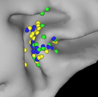
This image shows the locations of single-units recorded during an oculomotor delayed response task in the frontal eye fields (FEF) of two macaque monkeys. In this image, FEF responses were classified based on the thee classical FEF response types: visual, motor, and visuomotor.
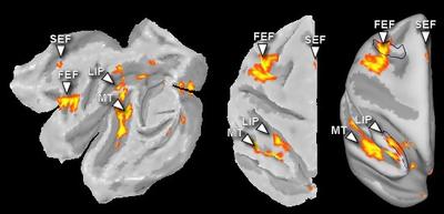
The positions of the frontal eye field (FEF), supplementary eye field (SEF), the lateral intraparietal area (LIP), and the middle temporal (MT) area on the macaque cortical surface, as determined by functional MRI during saccade execution.